ACL injury in Nagpur
ACL Injury: Understanding the Anatomy, Causes, Diagnosis, and Treatment
An injury to the Anterior Cruciate Ligament (ACL) is among the most prevalent knee injuries, especially in athletes. The ACL is essential for ensuring stability in the knee joint, and any injury to this ligament can greatly affect both mobility and athletic performance. This blog will examine the structure of the ACL, the mechanisms through which injuries happen, the diagnostic processes involved, and the treatment options available for recovery..
Anatomy of the ACL
The anterior cruciate ligament (ACL) is one of the four main ligaments in the knee, connecting the femur (thigh bone) to the tibia (shinbone). It is positioned diagonally at the center of the knee and is crucial for controlling the forward and backward movement of the tibia relative to the femur. The ACL’s primary purpose is to prevent excessive forward displacement of the tibia, thereby ensuring stability in the knee during activities such as walking, running, and jumping.
While the knee joint functions as a hinge joint, the ACL also contributes to the restriction of rotational movements that could potentially harm the knee. Consequently, it is essential for sports that require abrupt stops, directional changes, or jumping.
Description of ACL Injury
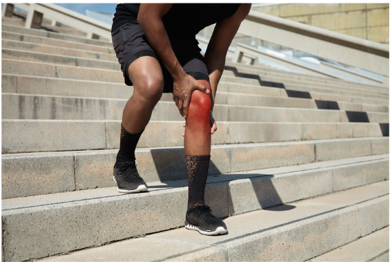
An injury to the anterior cruciate ligament (ACL) arises when the ligament is either overstretched or ruptured. Such injuries frequently occur in contact sports, including football, basketball, soccer, and skiing. The extent of the tear can vary, being either partial or complete, with a complete tear resulting in considerable instability within the knee joint.
Symptoms of an ACL injury
Symptoms can vary, but typical signs include:
- A pronounced “pop” noise occurring at the moment of injury
- Sudden pain experienced in the knee
- Rapid onset of swelling
- Inability to bear weight on the injured leg
- Restricted range of motion, especially when flexing or extending the knee
- A sensation of instability, as if the knee could collapse
An ACL injury can occur through multiple mechanisms, frequently resulting from a rapid halt, pivoting, or alteration in direction. Additionally, a direct impact to the knee from the side or front during a collision may also lead to a tear.
Causes of ACL Injury
ACL injuries are frequently linked to high-impact sports or activities characterized by abrupt stops, directional changes, or jumping.
Such injuries may arise in numerous situations:
- Rotational or Twisting Movements: When an athlete swiftly alters their direction, particularly with a foot firmly planted, it can place significant strain on the ACL, potentially leading to a tear.
- Jumping and Landing: The impact experienced upon landing from a jump, especially if the knee is twisted or hyperextended, can result in ACL damage.
- Direct Impact: A forceful strike to the knee or leg can lead to an ACL tear, a common occurrence in contact sports such as football or soccer.
- Overextension: If the knee joint is compelled to extend beyond its typical range of motion, such as during a fall, it may sustain an ACL tear.
- Muscle Weakness or Inadequate Technique: Insufficient conditioning, strength, or technique can elevate the risk of ACL injuries. Weakness in the quadriceps, hamstrings, or hip muscles, along with improper posture or alignment, can further exacerbate the likelihood of injury.
Diagnosis of ACL Injury
The assessment of an ACL injury is usually determined through a combination of physical examination and imaging studies. The following outlines the typical approach that medical professionals take when diagnosing an ACL injury:
- Physical Examination: The physician will assess the knee for indications of swelling, instability, and tenderness. They may conduct specific assessments, such as the Lachman test or the Anterior Drawer test, to evaluate for abnormal movement or looseness in the knee, which may suggest ligament injury.
- Imaging Tests:
- X-rays: Although X-rays are not capable of revealing soft tissue injuries like ligament tears, they are utilized to exclude the possibility of bone fractures that may accompany an ACL injury.
- MRI (Magnetic Resonance Imaging): An MRI is considered the most precise imaging modality for diagnosing an ACL tear. It offers detailed images of soft tissues, enabling physicians to ascertain the severity of the injury and eliminate the possibility of damage to other knee components, such as the meniscus.
- Arthroscopy: In certain situations, a surgeon may carry out an arthroscopic evaluation, wherein a small camera is inserted into the knee joint. This procedure provides a direct view of the injury, assisting in both diagnosis and treatment.
Treatment for ACL Injury
The management of an ACL injury is contingent upon the extent of the tear, the age of the patient, their level of physical activity, and their general health status. There are two main strategies for treatment: conservative (non-surgical) management and surgical procedures.
Non-Surgical Treatment:For individuals with partial ACL tears or those who prefer not to resume high-demand sports, non-surgical treatment may be considered a viable alternative. This approach generally includes:
- Physical Therapy: Engaging in a rehabilitation program is crucial for fortifying the muscles surrounding the knee and enhancing the range of motion. The exercises primarily target the quadriceps, hamstrings, and stabilizing muscles to offset the effects of the injured ACL.
- Bracing: The use of a knee brace can offer supplementary support and stability to the knee during physical activities.
- Medication: Non-steroidal anti-inflammatory drugs (NSAIDs) may be recommended to alleviate pain and reduce inflammation.
Surgical Treatment:
In cases where the ACL tear is significant or the individual desires to resume sports or activities that require high physical exertion, surgical intervention is frequently required. The predominant surgical approach is ACL reconstruction, which entails:
- Autograft versus Allograft: A tendon may be sourced from the patient’s own body (autograft) or from a donor (allograft) to replace the damaged anterior cruciate ligament (ACL). The most frequently utilized tendons include the patellar tendon, hamstring tendon, and quadriceps tendon.
- Arthroscopic Surgery: This technique is characterized by its minimally invasive nature, employing small incisions and a camera to assist the surgeon in the repair or reconstruction of the ligament.
- Post-Surgical Rehabilitation: Following the surgical procedure, engaging in physical therapy is essential for regaining strength and flexibility in the knee. Recovery durations can differ significantly, typically spanning from six months to a year, contingent upon the individual’s circumstances and activity level.
Recovery and Prevention
Rehabilitation is essential for achieving a complete recovery, irrespective of the treatment approach employed. Throughout the recovery process, emphasis should be placed on strength training, flexibility, and proprioception, which encompasses balance and coordination. A tailored rehabilitation program is crucial in facilitating the knee’s return to optimal functionality.
To prevent future ACL injuries, athletes and individuals should:
- Enhance the musculature surrounding the knee: Well-developed quadriceps, hamstrings, and calf muscles offer improved support for the anterior cruciate ligament (ACL).
- Utilize correct techniques: Athletes must be trained to land appropriately and to steer clear of excessive twisting or hyperextension.
- Engage in adequate warm-up routines: Proper stretching and warm-up exercises prior to physical activity can diminish the likelihood of injury.
- Select suitable footwear: Supportive shoes can help mitigate the risk of twisting or other movements that may compromise the integrity of the ACL.